Effective Use of the Coloring Resource

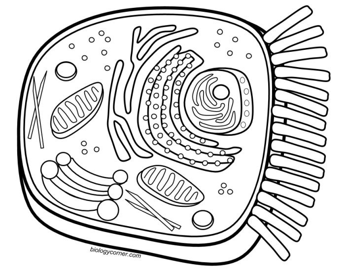
The “Biology Corner Animal Cell Coloring” resource provides a valuable hands-on activity for students learning about the intricate world of animal cells. Coloring engages different learning styles and helps solidify understanding of organelle structure and function. By incorporating this resource strategically, teachers can enhance their lesson plans and create a more memorable learning experience.This section explores several methods for effectively integrating the coloring page into lesson plans and assessment strategies.
It provides practical examples and activities to maximize student engagement and comprehension of animal cell biology.
Integrating the Coloring Page into Lesson Plans
The coloring page can be used in various stages of a lesson plan, from introduction to reinforcement. Introducing the coloring page after an initial lecture or reading assignment can help students visualize the organelles they have just learned about. It can also serve as a pre-teaching activity to spark curiosity and prior knowledge before diving into the complexities of cell structure.
Using the coloring page as a follow-up activity allows students to review and consolidate their learning.
Designing an Activity to Reinforce Learning
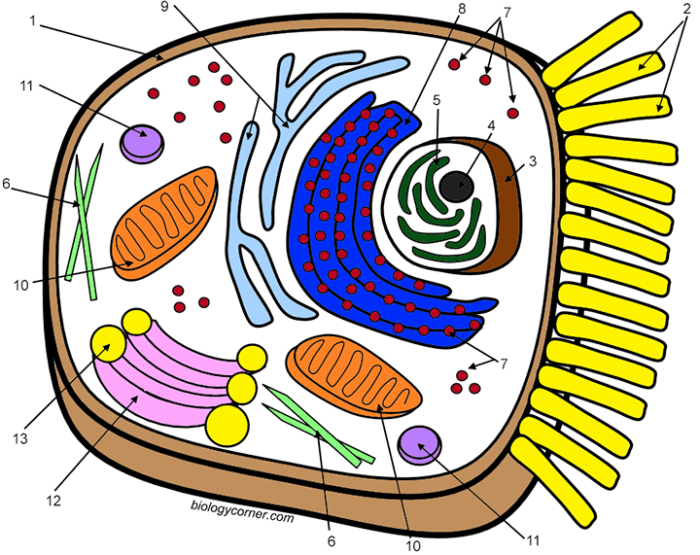
A structured activity can maximize the learning potential of the coloring page. One effective approach is to assign each organelle a specific color, connecting the color to the organelle’s function. For example, the mitochondria, the powerhouse of the cell, could be colored red to represent energy. Lysosomes, responsible for waste breakdown, could be colored brown, resembling waste material.
This color-coding strategy helps students visually connect structure and function. Furthermore, requiring students to label each organelle directly on the coloring page reinforces their understanding of organelle names and locations. A short presentation where students explain their color choices and the function of each organelle can further solidify their understanding and encourage peer learning.
Assessing Student Understanding, Biology corner animal cell coloring
The coloring activity provides several opportunities for assessment. The completed coloring page itself serves as a visual representation of student understanding. Accurate coloring and labeling demonstrate basic knowledge of organelle identification and location. A simple quiz based on the coloring page can assess factual recall. More in-depth assessment can be achieved by asking students to explain the function of each organelle in their own words, either in writing or during a class discussion.
For example, a student might explain that the Golgi apparatus is like the cell’s post office, packaging and distributing proteins. These explanations provide insight into the student’s deeper understanding of cellular processes. Alternatively, students can create a short story or comic strip featuring the organelles as characters, demonstrating their understanding in a creative and engaging way.
Visualizing Animal Cells: Biology Corner Animal Cell Coloring

Understanding the structure of an animal cell is fundamental to biology. This section aims to provide a clear visual representation of a typical animal cell, highlighting the appearance and location of its key organelles, and comparing this representation with what one might observe under a microscope. Furthermore, we’ll explore how color can be strategically employed in the coloring activity to emphasize the distinct roles of these organelles.
Imagine a rounded, somewhat irregular shape, like a fried egg with the yolk removed. This represents the basic form of an animal cell. The outer boundary is the cell membrane, a flexible barrier controlling what enters and exits the cell. Inside, a jelly-like substance called cytoplasm fills the cell, housing various organelles.
Appearance and Location of Key Organelles
Near the center of the cell resides the nucleus, a generally spherical body often depicted as darker than the surrounding cytoplasm. The nucleus contains the cell’s genetic material (DNA). Surrounding the nucleus is the endoplasmic reticulum (ER), a network of interconnected membranes. The rough ER, studded with ribosomes, appears slightly textured. Ribosomes, tiny structures scattered throughout the cytoplasm and on the rough ER, are responsible for protein synthesis.
The smooth ER, lacking ribosomes, appears smoother and is involved in lipid synthesis and detoxification. The Golgi apparatus, a stack of flattened, membrane-bound sacs, is typically located near the nucleus and plays a role in processing and packaging proteins. Mitochondria, the powerhouses of the cell, are oval-shaped organelles with a double membrane, the inner one folded into cristae.
Lysosomes, small spherical organelles, contain enzymes for breaking down waste materials. Centrioles, a pair of short, cylindrical structures, are involved in cell division.
Comparing Coloring Page and Microscopic Image
A coloring page provides a simplified and stylized representation of an animal cell, emphasizing the key organelles and their general arrangement. It’s like a map, highlighting the important landmarks. A microscopic image, on the other hand, shows a real cell with all its complexity. Organelles might appear less distinct, and their shapes can vary depending on the cell type and the microscopic technique used.
For example, the ER might appear as a blurry network rather than the distinct lines seen in a coloring page. The colors also differ. Microscopic images often use artificial colors to highlight specific structures, while a coloring page allows for more creative freedom.
Using Color to Highlight Organelle Function
Color can be a powerful tool for understanding organelle function in the coloring activity. For instance:
| Organelle | Suggested Color | Reason |
|---|---|---|
| Nucleus | Purple | Represents the importance of genetic information |
| Ribosomes | Blue | Links to the idea of protein synthesis as a building process |
| Mitochondria | Red/Orange | Reflects their role as energy producers |
| Lysosomes | Green | Associates with the breakdown and recycling of waste |
| Golgi Apparatus | Yellow | Highlights its role in processing and packaging |
| Endoplasmic Reticulum | Pink | Connects with the network-like structure |
This color-coding helps visualize the different roles each organelle plays within the cell.
Exploring the intricacies of cells with the ‘biology corner animal cell coloring’ activity provides a foundational understanding of life’s building blocks. For a delightful shift towards macroscopic life forms, explore these adorable animal coloring pages showcasing a variety of creatures. This artistic detour complements the cellular focus of the biology corner exercise by visualizing the complex organisms those cells create.


