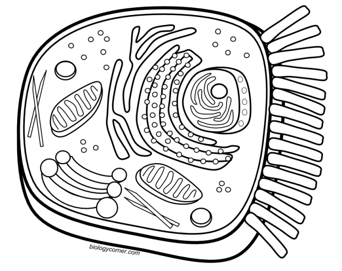
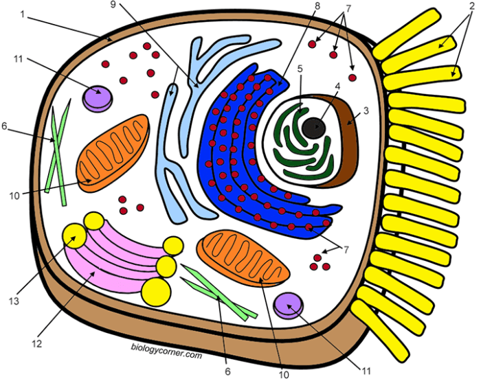
Target Audience for Animal Cell Coloring Sheets

Animal cell coloring sheets serve as a valuable educational tool for visualizing and understanding the complex structures within animal cells. They offer a hands-on learning experience, making the study of cell biology more engaging and accessible to various learners.Coloring sheets cater to a diverse range of learners, primarily within educational settings. The complexity of the diagrams and the accompanying information typically determine the appropriateness for different age groups and learning objectives.
Age Groups and Learning Objectives
Animal cell coloring sheets are adaptable to various age groups, each with specific learning objectives.
- Elementary School (Ages 6-10): At this level, coloring sheets introduce basic cell components like the nucleus, cell membrane, and cytoplasm. The focus is on visual recognition and associating these components with their basic functions. For example, students might learn that the nucleus is the “control center” of the cell. Coloring helps reinforce these initial concepts.
- Middle School (Ages 11-13): More detailed diagrams are introduced, featuring organelles like mitochondria, ribosomes, and the endoplasmic reticulum. Students learn about the specific roles of these organelles in cell processes, such as energy production and protein synthesis. Coloring activities can be combined with labeling exercises to reinforce the connection between structure and function.
- High School (Ages 14-18): Coloring sheets at this level can depict more complex cellular processes, like cell division or protein transport. These visuals support advanced coursework in biology, allowing students to visualize intricate interactions within the cell. The coloring activity itself might become less of a focus, serving more as a supplementary tool for understanding complex concepts.
Educational Settings, Animal cell coloring sheet
Animal cell coloring sheets find widespread use in a variety of educational contexts.
- Formal Classrooms: Teachers utilize coloring sheets as a supplementary activity to textbook learning, providing a visual and kinesthetic learning experience. They can be incorporated into lesson plans on cell biology, allowing students to actively engage with the material.
- Homeschooling Environments: Coloring sheets offer a flexible and engaging learning tool for homeschooling parents. They provide a hands-on activity that complements other educational resources and caters to different learning styles.
- Informal Learning Centers: Museums, science centers, and after-school programs often use coloring sheets as an interactive activity to introduce children to scientific concepts in an informal and engaging way.
Educational Value of Animal Cell Coloring Sheets
Animal cell coloring sheets offer a dynamic and engaging approach to learning about cell biology. They transform the complex world of cellular structures into a visually accessible and interactive experience, benefiting learners of all ages. By actively participating in coloring, students move beyond passive memorization and develop a deeper understanding of the intricate workings of animal cells.Coloring sheets bridge the gap between abstract concepts and tangible learning.
The act of coloring encourages focused attention on individual cell components, their shapes, and their relative positions within the cell. This focused interaction reinforces visual memory, making it easier to recall and identify these structures later.
Reinforcement of Cell Component Memorization
The process of coloring necessitates a close examination of each organelle’s structure and location within the cell. As students select colors for the nucleus, mitochondria, ribosomes, and other organelles, they are actively engaging with the material. This active engagement strengthens memory traces, leading to improved recall of cell components and their functions. Furthermore, associating specific colors with different organelles can create a mnemonic device, further aiding memorization.
Integration into Broader Lesson Plans
Animal cell coloring sheets can be seamlessly integrated into various stages of a cell biology lesson. They can serve as an introductory activity to spark curiosity and provide a visual overview of cell structure before diving into more complex concepts. During the lesson, coloring sheets can be used to reinforce newly learned information and provide a hands-on activity to solidify understanding.
As a follow-up activity, they can be used to assess comprehension and identify areas where students may need further clarification. For example, after a lecture on the functions of different organelles, students can color-code the organelles based on their roles (e.g., energy production, protein synthesis). This reinforces the connection between structure and function.
| Activity | Integration of Coloring Sheet |
|---|---|
| Introduction | Students color a basic animal cell, labeling each organelle as it is introduced. |
| Lesson Development | Students color specific organelles as their functions are discussed, using different colors to represent different processes. |
| Assessment | Students complete a coloring sheet with unlabeled organelles, demonstrating their ability to identify and differentiate each component. |
Common Components Featured on Animal Cell Coloring Sheets
Animal cell coloring sheets typically focus on the key organelles that contribute to the cell’s structure and function. These visual aids help learners identify and understand the roles of these components within the complex environment of the cell. Coloring the different organelles can enhance memory and comprehension of their distinct characteristics.
Organelles and Their Functions
The following table Artikels the common organelles featured on animal cell coloring sheets, along with their descriptions and primary functions. These organelles work together in a coordinated manner to maintain the cell’s life and carry out its specific tasks.
| Organelle | Description | Primary Function |
|---|---|---|
| Cell Membrane | A flexible outer boundary that controls the movement of substances in and out of the cell. | Protection, regulation of transport. |
| Cytoplasm | The jelly-like substance that fills the cell, containing the organelles. | Suspends organelles, site of many chemical reactions. |
| Nucleus | The control center of the cell, containing the cell’s genetic material (DNA). | Stores DNA, controls cell activities. |
| Mitochondria | The “powerhouses” of the cell, responsible for energy production. | Generates ATP (cellular energy) through respiration. |
| Ribosomes | Small structures responsible for protein synthesis. | Synthesizes proteins. |
| Endoplasmic Reticulum (ER) | A network of membranes involved in protein and lipid synthesis and transport. There are two types: Rough ER (with ribosomes) and Smooth ER (without ribosomes). | Protein and lipid synthesis, transport of molecules. |
| Golgi Apparatus | Processes, packages, and distributes proteins and lipids. | Modifies, sorts, and packages molecules for transport. |
| Lysosomes | Membrane-bound sacs containing enzymes that break down waste materials and cellular debris. | Digestion of waste materials, recycling of cellular components. |
| Vacuoles | Storage sacs for water, nutrients, and waste products. Animal cells typically have smaller vacuoles than plant cells. | Storage of water, nutrients, and waste. |
Accessibility and Variations of Animal Cell Coloring Sheets
Adapting educational materials to cater to diverse learning styles and needs is crucial for inclusive education. Animal cell coloring sheets, while visually engaging for many, can present challenges for students with visual impairments or other learning differences. Creating accessible versions of these resources ensures that all students can participate in learning about cell biology.Making animal cell coloring sheets accessible involves considering various adaptations and alternative formats.
These modifications aim to provide equitable learning opportunities for all students, regardless of their abilities.
Tactile and Raised-Line Animal Cell Diagrams
For students with visual impairments, tactile or raised-line diagrams offer a hands-on approach to understanding cell structure. These diagrams can be created using various materials, such as textured paper, puffy paint, or thermoform. Key cell components are represented by raised lines or different textures, allowing students to explore the cell’s organization through touch. For example, the nucleus could be represented by a rough circular patch, the endoplasmic reticulum by a series of raised wavy lines, and the ribosomes by small, raised dots.
Exploring animal cell coloring sheets can be a fun educational activity. For a wider range of animal visuals, check out these coloring book animals printable options for inspiration. Returning to the cellular level, understanding the structure of organelles within an animal cell becomes easier with a well-labeled coloring sheet.
These tactile representations provide a concrete way for visually impaired students to grasp the spatial relationships between different organelles.
Simplified Animal Cell Coloring Sheets with Large Print
Students with low vision may benefit from simplified coloring sheets with larger print and high contrast. These sheets feature fewer details and larger labels, making them easier to see and color. Using bold, black Artikels for the cell and its components against a light background enhances visibility. Reducing the number of organelles to focus on the main structures, like the nucleus, cytoplasm, cell membrane, and mitochondria, can also simplify the visual information and make it more accessible.
Digital Animal Cell Coloring Sheets with Audio Descriptions
Digital versions of animal cell coloring sheets offer further accessibility options. These can incorporate audio descriptions of the different cell components, providing auditory information for students with visual impairments. Interactive features, such as clickable organelles that provide further details or pronunciations, can enhance engagement and learning for all students. For example, clicking on the mitochondria could trigger a short audio clip explaining its function as the “powerhouse” of the cell.
Braille or Large Print Labels for Cell Components
Adding Braille or large print labels to existing animal cell coloring sheets can make them more inclusive for students with visual impairments or reading difficulties. These labels can be affixed directly to the coloring sheet or provided as a separate key. Clear, concise descriptions in Braille or large print allow students to independently identify and understand the different parts of the cell.
This adaptation enables them to participate in the coloring activity while simultaneously learning the names and functions of the organelles.
Integrating Animal Cell Coloring Sheets into Activities
Animal cell coloring sheets offer a dynamic and engaging approach to learning about cell biology. They transform the complex world of cellular structures into a hands-on activity, making learning more accessible and enjoyable for students of all ages. By combining visual learning with kinesthetic activity, coloring sheets cater to diverse learning styles and promote better retention of information.Coloring sheets can be seamlessly integrated into various classroom activities to reinforce learning and create a multi-faceted educational experience.
This approach helps bridge the gap between theoretical knowledge and practical application, allowing students to visualize and internalize the intricacies of animal cells.
Engaging Classroom Activity
A highly engaging classroom activity involves dividing students into small groups and assigning each group a specific organelle to research. After their research, each group creates a presentation about their assigned organelle, highlighting its function and importance within the cell. Following the presentations, students use colored pencils or markers to color their animal cell diagrams, paying special attention to the organelle they researched.
This activity fosters collaboration, research skills, and a deeper understanding of individual organelles and their roles within the cell.
Lesson Plan Incorporating Coloring Sheets
A comprehensive lesson plan incorporating animal cell coloring sheets can be structured across multiple stages. The lesson begins with an introductory discussion about cells as the basic units of life. A short video or interactive presentation can be used to introduce the different organelles and their functions. Then, students receive their animal cell coloring sheets and are guided through the different organelles, their shapes, and their locations within the cell.
After the coloring activity, students engage in a labeling exercise to reinforce their understanding of organelle names and positions. Finally, a class discussion or quiz can be used to assess learning and address any remaining questions.
Pre and Post-Coloring Activities
Pre-coloring activities can prepare students for a more meaningful coloring experience. For instance, a brainstorming session on cell functions can activate prior knowledge and spark curiosity. Alternatively, a quick review of cell vocabulary can equip students with the necessary terminology. Post-coloring activities can further solidify learning and assess comprehension. A “cell analogy” activity, where students compare organelles to real-world objects and their functions, can enhance understanding and critical thinking.
Another effective post-coloring activity involves having students create a 3D model of an animal cell using craft materials, further solidifying their understanding of spatial relationships within the cell.
Creating Your Own Animal Cell Coloring Sheet
Designing a custom animal cell coloring sheet allows for tailored learning experiences. This section Artikels the process of creating such a resource, from initial concept to final design, emphasizing best practices for visual clarity and educational effectiveness.Creating a personalized animal cell coloring sheet involves several key steps to ensure both accuracy and engagement. The following process guides you through building a coloring sheet from scratch.
Step-by-Step Creation Process
Creating an animal cell coloring sheet involves a structured approach.
- Start with a basic Artikel of the cell: Draw a large, irregular circle or oval to represent the cell membrane.
- Add the nucleus: Draw a smaller circle within the cell, slightly off-center, to represent the nucleus. Include a smaller, darkened circle within the nucleus for the nucleolus.
- Incorporate key organelles: Draw simplified representations of other important organelles, such as the mitochondria (oval shapes with inner folds), endoplasmic reticulum (a network of interconnected lines), Golgi apparatus (a stack of flattened sacs), ribosomes (small dots scattered throughout the cytoplasm), and lysosomes (small circles). Refer to a biology textbook or reputable online resource for accurate depictions.
- Label each organelle clearly: Use concise labels placed near each organelle. Consider using a legend or key if space is limited.
- Refine and finalize: Review the overall layout, ensuring clear visuals and accurate labeling. Adjust the sizes and positions of elements as needed to achieve a balanced and understandable representation.
Tips for Visuals and Labeling
Effective visuals and clear labeling enhance the educational value of the coloring sheet.Choosing appropriate visuals and labeling conventions ensures the clarity and educational value of the coloring sheet. Simple, recognizable shapes are best for representing organelles. Labels should be clear, concise, and positioned close to the corresponding organelle. A consistent font size and style contributes to readability.
Using Digital Tools for Design
Digital tools offer advanced design capabilities and ease of editing.Software programs like Adobe Illustrator, Inkscape (a free, open-source alternative), or even simpler drawing tools like Google Drawings can be utilized to create professional-looking animal cell coloring sheets. These tools allow for precise shapes, clean lines, and easy adjustments. For example, using layers in a program like Illustrator allows you to separate the Artikel of the cell from the labels, making edits and revisions simpler.
You can also easily export your finished design in various formats suitable for printing.
Beyond Coloring

A completed animal cell coloring sheet serves as a springboard for deeper exploration of cell biology. The act of coloring helps students visualize the different organelles and their arrangement within the cell, creating a foundational understanding. Building upon this visual foundation, various activities can solidify knowledge and encourage a more interactive learning experience.The following activities extend learning beyond the coloring sheet, promoting critical thinking and a deeper understanding of cellular structure and function.
Labeling the Animal Cell Diagram
After coloring, labeling the different organelles reinforces the connection between visual representation and nomenclature. This activity strengthens memory recall and helps students associate the organelle’s name with its appearance and location within the cell. Providing a word bank or a completed diagram as a reference can assist students in this process.
Creating 3D Animal Cell Models
Transforming a 2D coloring sheet into a 3D model brings the animal cell to life. Students can use various materials like clay, construction paper, or even edible materials to represent the different organelles. This hands-on activity encourages creativity and allows students to visualize the cell’s three-dimensional structure, reinforcing the spatial relationships between organelles. For example, the nucleus could be represented by a large gumdrop, the endoplasmic reticulum by folded ribbon, and the mitochondria by small, oval-shaped candies.
Writing Descriptive Paragraphs About Organelles
Writing descriptive paragraphs about individual organelles or the cell as a whole encourages deeper engagement with the material. Students can research the specific function of each organelle and describe its role within the cell. This activity promotes analytical thinking and strengthens writing skills while reinforcing biological concepts. For example, a student might describe the mitochondria as the “powerhouse of the cell,” explaining its role in producing energy through cellular respiration.
Follow-Up Activity: Animal Cell Organelle Functions Matching
This activity uses the labeled parts of the animal cell to further reinforce understanding of organelle function. Prepare a set of cards, each featuring the name of an organelle on one card and its corresponding function on another. Students then match the organelle name with its function. This activity reinforces the connection between structure and function within the animal cell.
For instance, a student would match the card labeled “Ribosome” with the card describing “protein synthesis.”


