Introduction to Animal Cell Structure

Animal cell coloring pdf answer key – The animal cell, a bustling metropolis of microscopic activity, is the fundamental building block of animal life. Unlike its plant counterpart, it lacks a rigid cell wall, allowing for a greater degree of flexibility and movement. Within its membrane-bound confines, a complex array of organelles work in concert to maintain life, carrying out essential processes from energy production to waste disposal.
Understanding the structure and function of these organelles is key to grasping the intricacies of animal biology.
Animal cells, while diverse in their specific functions depending on the organism and tissue type, share a common set of essential components. These organelles, each with a specialized role, contribute to the overall health and functioning of the cell. Their coordinated actions ensure the cell’s survival and its contribution to the larger organism.
Animal Cell Organelles and Their Functions
The following table provides a concise overview of the major organelles found in a typical animal cell, their structure, and their vital roles. Each organelle plays a distinct part in maintaining cellular homeostasis and carrying out the cell’s diverse functions.
| Organelle | Structure | Function | Image Description |
|---|---|---|---|
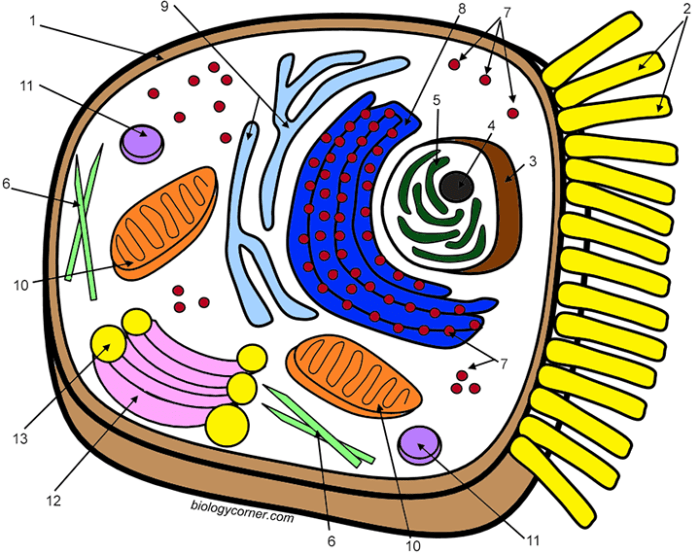
| Nucleus | Large, membrane-bound organelle containing the cell’s genetic material (DNA). | Controls cell activities, including growth, metabolism, and reproduction. Contains the genetic blueprint for the cell. | A large, round structure typically located near the center of the cell, often appearing darker than the surrounding cytoplasm. It may contain a visible nucleolus, a smaller, denser region involved in ribosome production. |
| Mitochondria | Double-membrane-bound organelles with folded inner membranes (cristae). | Powerhouses of the cell; responsible for cellular respiration, generating ATP (energy currency). | Rod-shaped or oval structures, often numerous within the cell. The inner membrane folds significantly, increasing surface area for ATP production. |
| Ribosomes | Small, granular structures, either free-floating in the cytoplasm or attached to the endoplasmic reticulum. | Protein synthesis; translate genetic information from mRNA into proteins. | Tiny dots scattered throughout the cytoplasm, sometimes appearing clustered near the endoplasmic reticulum. They are too small to be easily distinguished individually without high magnification. |
| Endoplasmic Reticulum (ER) | Network of interconnected membranes extending throughout the cytoplasm. Exists as rough ER (with ribosomes) and smooth ER (without ribosomes). | Rough ER: protein synthesis and modification. Smooth ER: lipid synthesis, detoxification, and calcium storage. | A complex network of interconnected tubules and sacs that appears as a maze-like structure within the cell. The rough ER has a rougher appearance due to the attached ribosomes. |
Differences Between Plant and Animal Cells
While both plant and animal cells are eukaryotic (possessing a membrane-bound nucleus), several key differences distinguish them. These differences reflect their distinct roles and adaptations within their respective organisms.
The most striking difference is the presence of a rigid cell wall in plant cells, composed primarily of cellulose. This provides structural support and protection, absent in the more flexible animal cells. Plant cells also contain chloroplasts, the sites of photosynthesis, enabling them to produce their own food using sunlight. In contrast, animal cells rely on consuming other organisms for energy.
Finally, plant cells typically have a large central vacuole, which plays a crucial role in maintaining turgor pressure and storing water and nutrients. Animal cells may have smaller vacuoles, or none at all.
Coloring Activities
Embark on a vibrant journey into the microscopic world! Preparing for your animal cell coloring adventure is as crucial as understanding the cell’s structure itself. A well-organized approach ensures an accurate and engaging learning experience, transforming a simple coloring exercise into a powerful tool for understanding complex biological concepts. Let’s gather our supplies and dive in!
The meticulous process of coloring an animal cell not only enhances understanding but also fosters artistic expression. By carefully coloring each organelle, you’re actively engaging with its function and location within the cell, strengthening your memory and comprehension. This hands-on activity bridges the gap between abstract diagrams and the tangible reality of cellular structures.
Materials Needed
To successfully color your animal cell, you’ll need a selection of high-quality art supplies. The right tools significantly impact the final result and the overall enjoyment of the process. Choosing vibrant colors that accurately represent the various organelles will enhance your understanding and create a visually appealing representation of the cell.
The essential materials include:
- A printed animal cell worksheet: This serves as your canvas, providing a detailed Artikel of the cell’s components.
- Colored pencils or markers: Opt for a range of colors to represent the different organelles accurately. For example, use a deep brown for the nucleus, a bright green for the chloroplast (if included), and various shades for the other organelles.
- A ruler (optional): This can be helpful for precise coloring and labeling, especially if your worksheet includes detailed diagrams.
- A pencil (optional): Lightly sketch in any additional details or labels before using colored pencils or markers to avoid mistakes.
- An eraser (optional): For correcting any mistakes made during the initial sketching phase.
Preparing the Coloring Worksheet, Animal cell coloring pdf answer key
Before you unleash your inner artist, prepare your worksheet strategically. This step is crucial for ensuring a smooth and accurate coloring experience. Proper preparation minimizes errors and allows for a more focused and enjoyable activity.
- Review the worksheet: Familiarize yourself with the labeled organelles and their functions. This preliminary step helps you visualize the final result and choose appropriate colors.
- Choose your color scheme: Select colors that not only accurately represent the organelles but also create a visually appealing image. Consider using a color key if your worksheet doesn’t provide one.
- Light sketching (optional): If desired, lightly sketch the boundaries of each organelle with a pencil before coloring. This aids in precise coloring and prevents color bleed.
- Begin coloring: Start with a light hand, building up color gradually. This allows for better control and avoids oversaturation.
- Labeling (optional): After coloring, label each organelle using a pencil or fine-tipped marker for added clarity and understanding.
Tips for Effective Organization
Organization is key to a successful coloring project. A methodical approach enhances accuracy and ensures a clear understanding of the cell’s structure.
Finding the answers for your animal cell coloring pdf can be challenging, especially for younger learners. A great way to bridge the gap between complex cell structures and engaging learning is to incorporate fun activities like those found on kids animal coloring pages , which can help familiarize them with animal forms before tackling the intricacies of cellular biology.
Returning to the animal cell coloring pdf, remember to focus on the key organelles and their functions for a complete understanding.
Here are some helpful tips:
- Work in sections: Color one organelle at a time to avoid confusion and ensure accuracy.
- Use a legend: Create a small color key to match colors with organelles for easy reference.
- Maintain clean lines: Avoid coloring outside the lines to ensure a neat and accurate representation.
- Take breaks: If you feel overwhelmed, take short breaks to avoid fatigue and maintain focus.
- Compare your work: After completing the coloring, compare your work to a diagram to check accuracy.
Common Animal Cell Coloring Worksheet Challenges

Embarking on the vibrant journey of coloring an animal cell can be surprisingly tricky! While seemingly a simple task, accurately depicting the intricate world within a cell requires careful attention to detail and a solid understanding of its components. Many students encounter common pitfalls that can hinder their understanding and lead to inaccurate representations. Let’s explore these challenges and learn how to overcome them.The accurate portrayal of an animal cell hinges on three key aspects: understanding the relative sizes of organelles, their precise locations within the cell’s boundaries, and the strategic use of color to differentiate these vital structures.
Failing to master these aspects can lead to a visually confusing and scientifically inaccurate representation.
Size and Location of Organelles
Correctly depicting the relative sizes and positions of organelles is crucial for a biologically accurate representation. Imagine trying to build a miniature city without knowing the scale of its buildings – some structures would be disproportionately large or small, and their placement would be chaotic. Similarly, if the nucleus is drawn too small compared to the cell’s overall size, or the mitochondria are scattered randomly instead of being concentrated in energy-demanding areas, the resulting image will lack accuracy.
For instance, the nucleus, the cell’s control center, should be prominently displayed, significantly larger than other organelles like ribosomes. The Golgi apparatus, responsible for packaging and transporting proteins, should be depicted near the endoplasmic reticulum, its primary supplier of materials. Understanding these spatial relationships is key to creating a realistic and scientifically sound image.
Effective Color Differentiation of Organelles
Color plays a vital role in distinguishing the various organelles within the cell. Using a monotonous palette can create a muddled and indistinguishable image. Think of it like a map – without different colors to represent different countries or terrains, navigation would be impossible. Similarly, using distinct colors for each organelle allows for easy identification and understanding.
For example, a vibrant green for the chloroplasts (if depicting a plant cell, which this exercise does not), a deep blue for the nucleus, and a bright orange for the mitochondria would create a visually clear and informative representation. This ensures that each organelle stands out and its function is easily identifiable through its unique color coding. The choice of color itself is not rigidly defined, but the crucial element is clear differentiation.
Common Mistakes in Animal Cell Coloring
Three common mistakes frequently observed in student work are: (1) inaccurate sizing of organelles, often leading to a disproportionate representation; (2) incorrect placement of organelles, which fails to reflect their functional relationships; and (3) the use of a limited color palette that hinders clear differentiation between organelles, making it difficult to identify each structure. Addressing these mistakes requires careful planning and a strong understanding of cell biology.
By understanding the relative sizes and locations of organelles and using color strategically, students can create accurate and visually appealing representations of animal cells.
Advanced Coloring Techniques for Enhanced Learning
Unlocking a deeper understanding of animal cell structure goes beyond simple coloring. By employing advanced coloring techniques, you can transform your worksheet from a static image into a dynamic, three-dimensional representation of a living cell, enhancing your comprehension and memory retention. These techniques move beyond simple flat coloring, allowing for a more nuanced and insightful study of the cell’s intricate components.
The following techniques, when applied thoughtfully, will significantly improve your visualization of the complex interplay of organelles within the animal cell.
Advanced Coloring Techniques
Employing these techniques transforms a simple coloring exercise into a powerful learning tool, fostering a deeper understanding of the cell’s structure and function. Each technique adds a layer of complexity, mirroring the intricate nature of the cell itself.
- Shading: Shading uses variations in color intensity to create depth and dimension. For example, you can use a lighter shade of blue for the cytoplasm in the foreground and gradually darken it towards the edges, giving the impression of a three-dimensional space. Similarly, you can use darker shades to indicate areas where organelles are layered or overlapping, such as the Golgi apparatus layered behind the endoplasmic reticulum.
This technique provides a sense of volume and helps distinguish between overlapping structures.
- Highlighting: Highlighting focuses on emphasizing specific structures and their relationships. Using bright, contrasting colors for key organelles, such as a vibrant yellow for the nucleus or a bright green for the mitochondria, can draw attention to their importance and location within the cell. This allows you to visually isolate and study individual components in the context of the entire cell.
- Texture: Adding texture simulates the surface characteristics of organelles. For instance, you could use fine lines or stippling to depict the rough endoplasmic reticulum’s ribosome-studded surface, differentiating it visually from the smooth endoplasmic reticulum. Similarly, you could use a slightly grainy texture for the cell membrane to visually represent its complex lipid bilayer structure. This adds a layer of realism, further enhancing the learning experience by making the cell appear more lifelike.
Assessing Understanding Through Coloring Activities: Animal Cell Coloring Pdf Answer Key
The vibrant hues of a meticulously colored animal cell diagram aren’t just aesthetically pleasing; they represent a student’s grasp of complex biological structures. Evaluating these coloring exercises goes beyond simply checking for correct colors; it’s about assessing their understanding of the cell’s components, their relative sizes and locations, and their overall interconnectedness. A well-designed assessment strategy ensures that the activity serves as a genuine learning tool, not just a busywork assignment.Assessing the accuracy and completeness of a student’s work involves a multi-faceted approach.
It requires careful observation of both the technical aspects – the correct coloring and labeling of organelles – and the conceptual understanding demonstrated through the organization and representation of the cell’s components. In essence, the coloring activity becomes a visual representation of their learning journey, offering insights into their strengths and areas needing further development.
Animal Cell Coloring Rubric
A rubric provides a structured framework for evaluating student work, ensuring consistent and fair grading. The following rubric uses a three-column layout to clearly present the assessment criteria, the rating scale, and the corresponding points for each criterion.
| Criteria | Rating Scale | Points |
|---|---|---|
| Accuracy of Organelle Identification and Placement | Excellent (All organelles correctly identified and positioned); Good (Most organelles correctly identified and positioned, minor errors); Fair (Several organelles incorrectly identified or positioned); Poor (Many organelles missing or incorrectly identified and positioned) | 4, 3, 2, 1 |
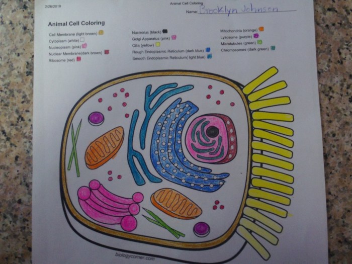
| Correctness of Organelle Coloring | Excellent (All organelles colored correctly according to the key); Good (Most organelles colored correctly, minor discrepancies); Fair (Several organelles incorrectly colored); Poor (Many organelles incorrectly colored or uncolored) | 4, 3, 2, 1 |
| Neatness and Organization | Excellent (Neat and organized coloring, clear labeling); Good (Mostly neat and organized, minor smudging or untidiness); Fair (Some areas messy or disorganized); Poor (Very messy and disorganized, difficult to interpret) | 3, 2, 1, 0 |
| Overall Completeness | Excellent (All required organelles included and labeled); Good (Most required organelles included and labeled); Fair (Several required organelles missing or unlabeled); Poor (Many required organelles missing or unlabeled) | 3, 2, 1, 0 |
Providing Constructive Feedback
Effective feedback is crucial for student learning. It should be specific, actionable, and focused on helping students improve their understanding. Instead of simply stating “incorrect,” teachers should pinpoint the exact error, such as “The Golgi apparatus should be located near the endoplasmic reticulum, not near the nucleus.” Similarly, instead of just saying “messy,” feedback might be, “Try using lighter strokes to avoid smudging, and consider outlining the organelles before coloring to improve organization.” Offering suggestions for improvement, such as “Review the diagrams in your textbook to reinforce the relative sizes and locations of the organelles,” fosters a growth mindset and encourages students to actively engage in their learning.
Positive reinforcement should also be incorporated, highlighting aspects of the work that are done well, even if areas need improvement. For example, “Your labeling is very clear and easy to read,” or “You’ve accurately depicted the cell membrane’s structure.” This balanced approach ensures that feedback is both supportive and instructive.
Q&A
Where can I find free printable animal cell coloring worksheets?
Many educational websites and online resources offer free printable animal cell coloring worksheets. A simple online search should yield numerous results.
What’s the best way to represent the cell membrane in my coloring?
Use a thin, continuous line to represent the cell membrane, differentiating it from the organelles inside. You can use a slightly darker shade to provide depth.
How important is accurate color representation in animal cell coloring?
While not strictly necessary for scientific accuracy, using different colors for different organelles helps in visual differentiation and improves learning and understanding.
Are there any online resources to check my answers after coloring?
While a specific online answer key might be hard to find, comparing your work to labeled diagrams and descriptions in textbooks or online resources will help verify accuracy.


