Introduction to Animal Cell Structures
Animal cell coloring key answers – Animal cells, the fundamental building blocks of animals, are complex and fascinating miniature factories, each performing a multitude of vital functions to sustain life. Understanding their intricate structures is key to grasping the mechanics of life itself. These cells, unlike their plant counterparts, lack a rigid cell wall and chloroplasts, leading to significant differences in their overall structure and function.The basic components of an animal cell work in a coordinated manner to maintain homeostasis and carry out the cell’s essential processes.
Need help with your animal cell coloring key answers? Understanding cell structures can be challenging, but a creative approach can help. For a fun break, try some intricate designs; check out these animal mandala coloring pages for a relaxing change of pace. Returning to your animal cell diagrams afterward, you might find the details clearer and easier to remember.
These components include the nucleus, cytoplasm, mitochondria, and the cell membrane, among many others. Each plays a unique and indispensable role in the cell’s overall operation.
The Nucleus: The Cell’s Control Center
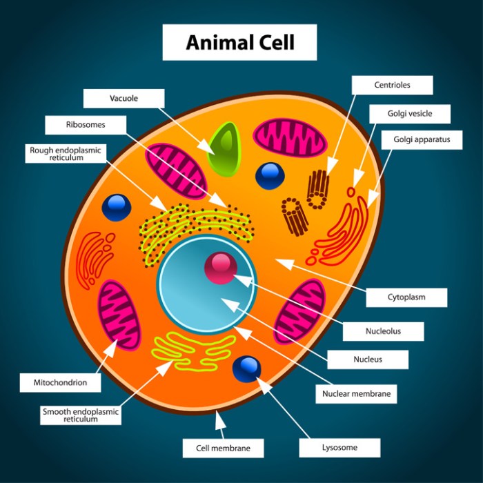
The nucleus is the cell’s central command center, housing the genetic material—DNA—organized into chromosomes. This DNA contains the blueprint for all cellular activities, dictating the synthesis of proteins and regulating the cell’s growth, division, and overall function. The nucleus is enclosed by a double membrane called the nuclear envelope, which is punctuated by nuclear pores that regulate the transport of molecules in and out of the nucleus.
Within the nucleus, a dense region called the nucleolus is responsible for the production of ribosomes, essential for protein synthesis. Imagine the nucleus as the brain of the cell, directing all operations.
The Cytoplasm: The Cell’s Internal Environment
The cytoplasm is the jelly-like substance filling the cell between the nucleus and the cell membrane. It’s a dynamic environment where many metabolic processes occur. Various organelles, including the mitochondria and ribosomes, are suspended within the cytoplasm. The cytoplasm provides a medium for the transport of molecules and facilitates cellular reactions. Think of the cytoplasm as the cell’s bustling workshop, where countless tasks are carried out simultaneously.
Mitochondria: The Powerhouses of the Cell
Mitochondria are often referred to as the “powerhouses” of the cell because they are responsible for generating most of the cell’s energy in the form of ATP (adenosine triphosphate) through cellular respiration. These bean-shaped organelles have their own DNA and ribosomes, suggesting an endosymbiotic origin. The inner membrane of the mitochondrion is highly folded, creating cristae that increase the surface area for ATP production.
The energy generated by mitochondria fuels all cellular activities, from muscle contraction to protein synthesis. Without them, the cell would be unable to function.
The Cell Membrane: The Cell’s Protective Barrier
The cell membrane, also known as the plasma membrane, forms the outer boundary of the animal cell. It’s a selectively permeable barrier that regulates the passage of substances into and out of the cell. This membrane is composed primarily of a phospholipid bilayer with embedded proteins. These proteins act as channels, carriers, or receptors, facilitating the transport of specific molecules.
The cell membrane maintains the cell’s internal environment, protecting it from external threats and controlling the flow of nutrients and waste products. It is a dynamic structure, constantly adjusting to the cell’s needs.
Animal Cells vs. Plant Cells: A Key Comparison
Animal and plant cells share some similarities, being both eukaryotic cells containing membrane-bound organelles. However, several key differences exist. Plant cells possess a rigid cell wall made of cellulose, providing structural support and protection, which is absent in animal cells. Plant cells also contain chloroplasts, the sites of photosynthesis, enabling them to produce their own food, unlike animal cells which are heterotrophic and rely on external sources of nutrients.
Furthermore, plant cells typically have a large central vacuole for storage and turgor pressure maintenance, while animal cells may have smaller vacuoles or none at all. These differences reflect the distinct lifestyles and adaptations of plants and animals.
Illustrating Cell Structures

Delving deeper into the animal cell, we now visualize the intricate architecture of its key components. Understanding their appearance is crucial to grasping their function within the dynamic cellular environment. The following descriptions will paint a vivid picture of these remarkable structures.
The detailed visualization of these structures, while challenging to fully capture without microscopy, is essential for a comprehensive understanding of their roles in cellular processes.
Endoplasmic Reticulum: Rough and Smooth
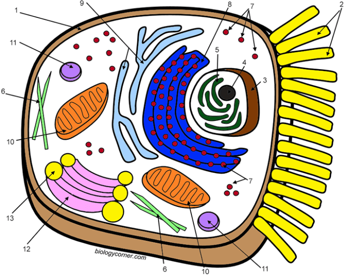
The endoplasmic reticulum (ER) is a vast network of interconnected membranes extending throughout the cytoplasm. Imagine a complex, folded sheet of fabric, constantly shifting and reforming. The rough ER, studded with ribosomes (appearing as small, dark granules under a microscope), is responsible for protein synthesis. These ribosomes give the rough ER its characteristic bumpy appearance. In contrast, the smooth ER lacks ribosomes and appears as a smoother, less textured network.
Its primary functions include lipid synthesis, detoxification, and calcium storage. The smooth ER’s appearance is more uniform and less granular compared to its rough counterpart. The difference in appearance directly reflects the difference in their primary functions: protein synthesis versus lipid metabolism.
Cell Membrane: The Fluid Mosaic Model
The cell membrane, the boundary of the cell, is not a static barrier but a dynamic, fluid structure. Imagine a sea of phospholipids, constantly moving and shifting. Within this sea are embedded various proteins, like icebergs floating in the ocean. These proteins have diverse functions, including transport, cell signaling, and cell adhesion. Carbohydrates are also attached to some of these proteins and lipids, contributing to the overall complexity of the membrane.
The fluid mosaic model emphasizes this constant movement and the variety of components within the membrane, creating a dynamic and ever-changing surface. The appearance, therefore, is not static; it’s a constantly fluctuating mosaic of lipids, proteins, and carbohydrates, giving it a visually complex, slightly blurry appearance under powerful microscopes.
Centrioles: Orchestrators of Cell Division
Centrioles are cylindrical structures, typically appearing as short, paired cylinders, situated near the nucleus. Imagine two tiny barrels lying side-by-side. Each centriole is composed of nine triplets of microtubules arranged in a circular pattern. These microtubules are protein fibers that provide structural support and are crucial for cell division. During cell division, centrioles duplicate and migrate to opposite poles of the cell, organizing the mitotic spindle, a complex structure that separates chromosomes into daughter cells.
The precise arrangement of the microtubules is essential for accurate chromosome segregation, ensuring that each daughter cell receives a complete set of genetic material. Without functional centrioles, cell division would be chaotic and likely unsuccessful. The visual representation would show these small, cylindrical structures near the nucleus, often radiating microtubules extending towards the cell periphery during cell division.
Coloring Activities and Exercises

Embark on a vibrant journey into the microscopic world by applying your newly acquired knowledge of animal cell structures through engaging coloring activities. These exercises will reinforce your understanding of cell components and their relative positions within the cell. Accurate coloring and labeling will solidify your grasp of this fundamental biological concept.This section provides a step-by-step guide for coloring and labeling animal cell diagrams, enhancing your comprehension through hands-on practice.
We’ll explore how to translate the information from your coloring key into a visually appealing and scientifically accurate representation of an animal cell.
Coloring an Animal Cell Diagram
Using your completed coloring key, meticulously color the provided animal cell diagram. Ensure each organelle is accurately represented by its assigned color. For example, the nucleus, designated as purple in your key, should be colored uniformly purple, with clear boundaries to distinguish it from the surrounding cytoplasm. Similarly, the mitochondria, perhaps colored a vibrant orange, should be depicted accurately as small, bean-shaped organelles scattered throughout the cytoplasm.
Pay close attention to detail; the clarity of your coloring will directly reflect your understanding of the cell’s structure. Consistent coloring and clear demarcation between organelles are essential for a successful and informative representation.
Creating a Labeled Animal Cell Diagram
Begin by drawing a circle to represent the cell membrane. Within this circle, sketch the various organelles, proportionally representing their sizes relative to one another. For instance, the nucleus, usually the largest organelle, should be centrally located and significantly larger than the mitochondria or ribosomes. Next, refer to your coloring key. Using the assigned colors, carefully color each organelle.
Finally, label each colored organelle using neat, legible handwriting or a fine-tipped marker. Ensure labels are clearly connected to their respective organelles using straight lines. This process not only enhances your understanding of the cell’s structure but also improves your scientific illustration skills.
Animal Cell Diagram Worksheet
The following represents a description of a worksheet containing several unlabeled animal cell diagrams. Each diagram provides a slightly different perspective or level of detail, allowing for varied practice. Diagram A, for example, might show a simplified cell with only the major organelles. Diagram B could be more complex, including smaller structures like ribosomes. Diagram C might present a cross-section view.
Students should utilize the provided coloring key to color and label each diagram accurately. This exercise reinforces their knowledge of animal cell structures through repetition and application. This repeated practice will solidify understanding and improve labeling accuracy.
Advanced Cell Components: Animal Cell Coloring Key Answers
Delving deeper into the intricate world of animal cells reveals a complex network of structures that orchestrate cellular function with remarkable precision. Beyond the readily identifiable organelles, a fascinating array of advanced components contributes to the cell’s dynamic equilibrium and overall vitality. Understanding these components provides a more complete picture of the cell’s remarkable capabilities.The cytoskeleton, a dynamic and versatile scaffolding system, is crucial for maintaining cell shape, facilitating intracellular transport, and enabling cell motility.
This intricate network comprises three primary components: microtubules, microfilaments, and intermediate filaments, each with unique structural properties and functions.
Cytoskeletal Components and Their Functions
Microtubules, the largest of the cytoskeletal filaments, are hollow tubes composed of the protein tubulin. Their primary roles include providing structural support, guiding intracellular transport via motor proteins like kinesin and dynein, and forming the mitotic spindle during cell division. Imagine them as the cell’s highways, directing the movement of organelles and vesicles. Microfilaments, composed of actin, are thinner and more flexible than microtubules.
They are involved in cell shape changes, muscle contraction (through interaction with myosin), and cytokinesis (the division of the cytoplasm during cell division). Picture them as the cell’s muscles, enabling movement and contraction. Intermediate filaments, as their name suggests, are intermediate in size between microtubules and microfilaments. They provide mechanical strength and structural support to the cell, anchoring organelles and resisting tensile forces.
They act as the cell’s strong internal ropes, preventing excessive stretching and damage.
Vesicle Diversity and Function
Cells utilize a diverse range of vesicles for various transport and storage purposes. These membrane-bound sacs vary in size, composition, and function. For instance, transport vesicles shuttle proteins and lipids between the endoplasmic reticulum, Golgi apparatus, and plasma membrane. Secretory vesicles store and release hormones or neurotransmitters. Lysosomes contain hydrolytic enzymes for waste breakdown and recycling.
Endosomes mediate endocytosis, the process of bringing materials into the cell. The specific contents and destination of a vesicle dictate its function within the complex cellular machinery. Consider a vesicle as a specialized delivery package, with its contents and destination clearly labelled.
Nuclear Envelope and Gene Expression Regulation, Animal cell coloring key answers
The nuclear envelope, a double membrane structure surrounding the nucleus, plays a vital role in regulating gene expression. Nuclear pores, embedded within the envelope, selectively control the passage of molecules between the nucleus and cytoplasm. This precise control ensures that only necessary molecules, such as RNA and regulatory proteins, can access the genetic material within the nucleus, while others are excluded.
The inner nuclear membrane is associated with the nuclear lamina, a protein meshwork that provides structural support to the nucleus and plays a role in gene organization and regulation. The intricate structure and selective permeability of the nuclear envelope are critical for maintaining the integrity of the genome and regulating gene expression. This sophisticated system ensures that genetic information is both protected and efficiently utilized.
FAQ Section
What are some common mistakes when using a coloring key for animal cells?
Common mistakes include misinterpreting the key’s color assignments, incorrectly labeling organelles, and failing to accurately represent the relative sizes and positions of organelles.
How can I improve my understanding of animal cell structures?
Utilize online resources, textbooks, and interactive simulations to further your knowledge. Building 3D models can also significantly enhance comprehension.
Are there different types of animal cells?
Yes, animal cells exhibit diversity in structure and function depending on their tissue type and role within the organism. Nerve cells, muscle cells, and epithelial cells are examples of specialized animal cells.
Where can I find additional coloring activities for animal cells?
Educational websites and biology textbooks often provide supplementary coloring activities and worksheets.


