Understanding Animal Cells
Animal cell coloring colored – Animal cells are the fundamental building blocks of all animals, from the simplest sponge to the most complex human. They are eukaryotic cells, meaning they possess a defined nucleus containing the cell’s genetic material. Unlike plant cells, animal cells lack a cell wall and chloroplasts.The basic structure of a typical animal cell includes a cell membrane, cytoplasm, and a nucleus.
The cell membrane, a selectively permeable barrier, encloses the cell’s contents and regulates the passage of substances in and out. The cytoplasm, a gel-like substance, fills the cell and houses various organelles. The nucleus, the control center of the cell, contains the cell’s DNA, which directs all cellular activities.
Exploring the intricate world of animal cell coloring, with its vibrant organelles and complex structures, can be fascinating. For a simpler, more whimsical take on animal imagery, check out these coloring pages cute animals which offer a fun alternative. Returning to the cellular level, accurately colored animal cell diagrams are invaluable educational tools for understanding biological processes.
Organelle Functions
Within the cytoplasm, numerous organelles perform specific functions essential for cell survival and function. The mitochondria, often referred to as the “powerhouses” of the cell, generate energy through cellular respiration. Ribosomes synthesize proteins, essential for cell structure and function. The endoplasmic reticulum (ER) is involved in protein and lipid synthesis, with the rough ER studded with ribosomes and the smooth ER lacking ribosomes.
The Golgi apparatus modifies, sorts, and packages proteins and lipids for transport within or outside the cell. Lysosomes contain enzymes that break down waste materials and cellular debris. The cytoskeleton provides structural support and facilitates cell movement. Centrioles play a role in cell division.
Specialized Animal Cell Examples
Animal cells exhibit remarkable diversity in structure and function, reflecting their specialized roles within the organism.
| Cell Type | Specialization |
|---|---|
| Nerve cells (neurons) | Transmit electrical signals throughout the body, enabling communication and coordination. They have long, branching extensions called axons and dendrites. |
| Muscle cells | Responsible for movement. They contain contractile proteins that allow them to shorten and generate force. Examples include skeletal muscle cells, smooth muscle cells, and cardiac muscle cells. |
| Red blood cells (erythrocytes) | Transport oxygen throughout the body. They are disc-shaped and lack a nucleus, maximizing their oxygen-carrying capacity. |
| White blood cells (leukocytes) | Part of the immune system, defending the body against pathogens. Different types of white blood cells, such as neutrophils and lymphocytes, have specialized roles in immune responses. |
| Epithelial cells | Form linings and coverings throughout the body, protecting underlying tissues and regulating the passage of substances. Examples include skin cells and the lining of the digestive tract. |
Visual Representations of Animal Cells

Visualizing the intricate world of animal cells is crucial for understanding their structure and function. Effective visual representations, especially those designed for interactive engagement like coloring, can significantly enhance comprehension of these complex biological units.
Detailed Illustration of an Animal Cell for Coloring
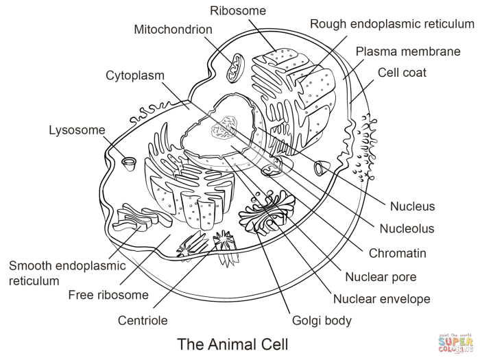
Imagine a circular or slightly irregular shape representing the cell membrane, the cell’s outer boundary. Within this boundary, the cytoplasm fills the space, depicted as a lighter shade. The nucleus, the cell’s control center, resides near the center, a prominent, darker circle containing a smaller, even darker nucleolus. Scattered throughout the cytoplasm are various organelles. Mitochondria, the powerhouses of the cell, are depicted as bean-shaped structures with internal folds.
The endoplasmic reticulum, a network of interconnected membranes, appears as a series of folded lines and tubes extending from the nucleus. Ribosomes, tiny dots scattered throughout the cytoplasm and on the endoplasmic reticulum, synthesize proteins. Golgi bodies, flattened sacs stacked together, process and package proteins. Lysosomes, small, spherical organelles, function as the cell’s recycling centers. Vacuoles, depicted as clear, membrane-bound sacs, store water and other materials.
Centrioles, a pair of small cylindrical structures near the nucleus, play a role in cell division. This illustration, with its clear Artikels and distinct representation of each organelle, provides a comprehensive visual of an animal cell.
Coloring Techniques for Enhancing Visual Representation, Animal cell coloring colored
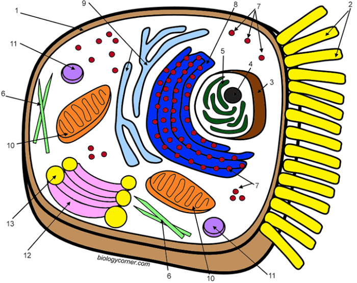
Different coloring techniques can highlight the distinct roles of each organelle. Using warm colors like reds and oranges for the mitochondria emphasizes their energy-producing function. Cooler blues and greens can represent the endoplasmic reticulum and Golgi bodies, reflecting their roles in protein synthesis and processing. Purple or deep blue can be used for the nucleus, signifying its importance as the control center.
Contrasting colors between the organelles and the cytoplasm create a clear distinction, enhancing visual clarity and aiding in memorization. Using varying shades within each organelle can also add depth and dimension, making the illustration more visually appealing.
Importance of Accurate and Engaging Visuals
Accurate and engaging visuals play a vital role in understanding complex biological structures like animal cells. These visuals translate abstract concepts into tangible representations, making them more accessible and easier to grasp. A well-designed illustration, particularly one that allows for interactive engagement through coloring, can significantly improve comprehension and retention of information. The act of coloring itself reinforces learning by requiring active participation and focus on the different components of the cell.
This active learning approach promotes a deeper understanding of the structure and function of each organelle and how they work together within the complex system of the animal cell.
Educational Resources for Animal Cell Biology

Exploring the intricate world of animal cells often begins with a visual understanding of their structure and components. Coloring activities offer a hands-on approach to learning, allowing for a deeper engagement with the material. This section focuses on available resources and activities that enhance the learning experience beyond basic coloring.
Printable Animal Cell Coloring Pages and Online Resources
Numerous resources, both online and offline, provide printable animal cell coloring pages. These resources cater to different learning levels, from elementary school to advanced biology courses.
- Biologycorner.com: This website offers a variety of free printable resources, including animal cell diagrams suitable for coloring and labeling.
- Supercoloring.com: This platform provides a selection of animal cell coloring pages with varying levels of detail, suitable for different age groups.
- Educational supply stores: Physical stores often carry workbooks and educational materials that include animal cell diagrams for coloring.
- Science textbooks: Many biology textbooks include simplified animal cell diagrams specifically designed for student labeling and coloring activities.
Complementary Educational Activities
Coloring pages serve as a foundation for more interactive learning experiences. Engaging in activities that build upon the visual representation strengthens understanding and retention.
| Activity | Description |
|---|---|
| Labeling Exercises | After coloring the cell, students label the different organelles, reinforcing their names and functions. This can be done directly on the printed page or using a separate answer key. |
| 3D Model Building | Constructing a 3D model of an animal cell, using clay, foam, or other materials, brings the 2D representation to life. Students can assign different colors to the organelles, mirroring their colored diagram. |
| Interactive Online Games | Several online platforms offer interactive games and quizzes that test knowledge of animal cell structure and function. These games can reinforce concepts learned through coloring and labeling activities. |
| Creating Flashcards | Students create flashcards with the name of the organelle on one side and its function and a small colored image on the other. This promotes active recall and reinforces visual association. |
Creating Interactive Learning Experiences
Interactive learning goes beyond passive observation and encourages active participation. Utilizing colored animal cell diagrams as a starting point, several activities can be implemented to enhance the learning process.
| Activity | Description |
|---|---|
| Organelle Function Presentations | Divide students into groups and assign each group a specific organelle. They then research and present its function, using their colored diagrams as visual aids. |
| Comparative Cell Studies | Compare and contrast animal cells with plant cells, highlighting the differences in organelles and their functions using colored diagrams of both cell types. |
| Cell Analogy Projects | Students develop analogies for the cell and its organelles, relating them to real-world objects or systems. For example, the Golgi apparatus could be compared to a post office, packaging and distributing materials. |
| Virtual Cell Exploration | Utilize online interactive cell models that allow students to explore the cell in 3D and learn about the functions of each organelle in a dynamic environment. |
The Impact of Color in Scientific Visualization: Animal Cell Coloring Colored
Color plays a crucial role in scientific visualization, particularly in the study of complex subjects like cell biology. It transforms bland, potentially confusing diagrams into engaging and informative visuals, aiding comprehension and retention of intricate details. The strategic use of color can highlight key structures, differentiate components, and illustrate processes within the cell, making the information more accessible and memorable for learners.Color enhances understanding and memorization of scientific concepts by appealing to our visual processing systems.
Our brains are wired to respond to color, making colored diagrams more stimulating and easier to process than monochrome representations. This increased engagement leads to improved information encoding and recall. Furthermore, color can be used to represent different properties or functionalities, adding another layer of information to the visualization.
Color’s Role in Cellular Diagrams
Comparing colored and black-and-white diagrams of animal cells reveals the significant impact color has on comprehension. In black-and-white diagrams, cellular structures can appear crowded and difficult to distinguish. For example, the nucleus, mitochondria, and endoplasmic reticulum might blend together, making it challenging to understand their individual roles and relationships. Conversely, colored diagrams can clearly delineate each organelle using distinct hues.
A common example is using shades of pink or purple for the nucleus, orange for the mitochondria (representing energy production), and blue for the endoplasmic reticulum. This color-coding makes it easier to identify, locate, and remember the specific functions of each organelle.
Psychological Effects of Color in Educational Materials
Color evokes psychological responses that can be leveraged in educational materials. Certain colors are associated with specific emotions or concepts. For example, blue is often associated with calmness and stability, while red can represent energy or danger. Green is frequently linked to growth and nature. These associations can be strategically employed in educational materials to create a more engaging and memorable learning experience.
For instance, using cool colors like blue and green for background elements can create a sense of calm and focus, while using warmer colors like red and orange to highlight key structures can draw attention and emphasize their importance. Consider a diagram of a cell undergoing mitosis. Using different colors for each phase (e.g., blue for prophase, green for metaphase, yellow for anaphase, and red for telophase) not only distinguishes the phases but also subtly implies the progression and energy changes throughout the process.
This application of color psychology can enhance understanding and retention of complex cellular processes.


