Key Structures of Animal Cells in Coloring Exercises

Animal cell coloring answers – Coloring exercises provide a dynamic way to learn about the intricate world of animal cells. By actively engaging with the material, students can better visualize and retain information about the various organelles and their respective functions within the cell. This interactive approach strengthens understanding of the complex interplay within these fundamental biological units.
Organelles in Animal Cell Coloring Exercises
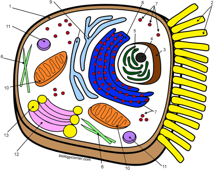
Typical animal cell coloring exercises feature a selection of key organelles crucial for cell function. Understanding these structures and their roles is fundamental to grasping the complexities of cellular biology.
- Cell Membrane
- Cytoplasm
- Nucleus
- Nucleolus
- Ribosomes
- Endoplasmic Reticulum (Smooth and Rough)
- Golgi Apparatus (or Golgi Body)
- Mitochondria
- Lysosomes
- Vacuoles
- Centrioles
Organelle Functions and Typical Coloring
The following table details the functions of the organelles commonly found in animal cell diagrams and suggests typical colors used in coloring exercises. These colors are suggestions and can be varied for individual preference or instructional purposes.
Understanding animal cell coloring answers can be complex, involving organelles and their functions. For a fun, simpler animal-related activity, check out this zoo animal coloring page featuring various creatures. Returning to cell diagrams, remember accurate coloring helps visualize and memorize each component’s role within the cell.
| Organelle | Function | Typical Coloring |
|---|---|---|
| Cell Membrane | Regulates the passage of substances into and out of the cell; provides structural support. | Light Pink/Purple |
| Cytoplasm | Jelly-like substance that fills the cell and surrounds the organelles; site of many metabolic reactions. | Light Yellow/Orange |
| Nucleus | Contains the cell’s genetic material (DNA) and controls cell activities. | Dark Purple/Blue |
| Nucleolus | Located within the nucleus; produces ribosomes. | Darker Purple/Blue than Nucleus |
| Ribosomes | Synthesize proteins. | Dark Blue/Black |
| Endoplasmic Reticulum (Smooth) | Synthesizes lipids, metabolizes carbohydrates, and detoxifies drugs and poisons. | Light Blue |
| Endoplasmic Reticulum (Rough) | Studded with ribosomes; involved in protein synthesis and modification. | Slightly Darker Light Blue |
| Golgi Apparatus (or Golgi Body) | Modifies, sorts, and packages proteins and lipids for storage or transport out of the cell. | Light Green/Tan |
| Mitochondria | Powerhouse of the cell; generates energy (ATP) through cellular respiration. | Orange/Red |
| Lysosomes | Contain digestive enzymes that break down waste materials and cellular debris. | Light Brown/Tan |
| Vacuoles | Store water, nutrients, and waste products. Generally smaller in animal cells than in plant cells. | Light Gray/Clear |
| Centrioles | Involved in cell division; help organize microtubules. | Dark Green/Brown |
The Role of Color in Visualizing Cellular Processes
Color acts as a powerful tool in representing complex biological processes, making them easier to understand and analyze. In the context of animal cells, color-coding helps distinguish various components and track their interactions during dynamic processes like cellular respiration and protein synthesis. By assigning specific colors to different molecules, organelles, or stages of a process, we can create visual narratives that enhance comprehension and facilitate communication of scientific information.Color-coding facilitates the illustration of dynamic processes within animal cells.
The strategic use of color can highlight the roles of different molecules and organelles as they interact and change throughout a cellular process. This visual aid simplifies the interpretation of complex mechanisms, making them more accessible to a wider audience.
Color Representation in Cellular Processes
Color can effectively represent different stages of cellular respiration or protein synthesis. For instance, in cellular respiration, the initial stage of glycolysis could be represented by a light blue, transitioning to a deeper blue as the process moves into the Krebs cycle. The final stage, the electron transport chain, could be depicted using shades of red to represent the increasing energy production.
Similarly, in protein synthesis, different colors could represent the various stages, such as transcription (green), mRNA processing (yellow), and translation (purple).
Visual Representation of Lysosomal Degradation
A visual representation of lysosomal degradation can effectively utilize color-coding to highlight its key steps. Imagine a circular cell depicted with a light gray cytoplasm. Within the cell, a lysosome is represented by a larger orange circle. A smaller, dark blue circle represents a damaged mitochondrion targeted for degradation.
1. Initial Contact
The blue mitochondrion approaches the orange lysosome. A light yellow halo surrounds the lysosome, representing the release of hydrolytic enzymes.
2. Engulfment
The orange lysosome begins to envelop the blue mitochondrion, with the blue gradually becoming encased within the orange. The yellow halo intensifies, indicating increased enzyme activity.
3. Degradation
The blue mitochondrion, now fully enclosed within the orange lysosome, begins to fragment. The blue color fades, replaced by smaller, lighter blue dots dispersed within the orange lysosome, representing the breakdown products.
4. Recycling
The orange lysosome, now containing the faded blue remnants, shows small green dots emerging from its surface. These green dots represent the recycled molecules being released back into the gray cytoplasm for reuse by the cell. The yellow halo diminishes, indicating a decrease in enzyme activity as the process concludes. This color-coded representation clearly illustrates the steps of lysosomal degradation, from initial contact to the recycling of components.
Educational Resources for Animal Cell Coloring: Animal Cell Coloring Answers

Coloring exercises offer a dynamic and engaging approach to learning about the intricate structures within animal cells. By actively participating in coloring activities, students can solidify their understanding of cellular components and their respective functions. This hands-on experience translates abstract concepts into visual representations, fostering deeper comprehension and retention.
Printable Animal Cell Coloring Sheets
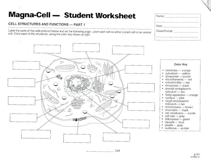
Numerous resources, both online and offline, offer printable animal cell coloring sheets suitable for various educational levels. Websites like Biology Corner, Education.com, and Pinterest provide a wide array of options, often categorized by complexity and detail. Offline resources include biology textbooks, workbooks, and educational supply stores. Selecting the right resource depends on the specific learning objectives and the student’s age and prior knowledge.
Benefits of Different Coloring Materials, Animal cell coloring answers
The choice of coloring materials can significantly impact the learning experience. Each medium offers distinct advantages in visualizing cellular structures and processes.
- Crayons: Ideal for younger learners, crayons provide a broad, vibrant color application, suitable for filling larger areas and emphasizing prominent structures like the nucleus and cell membrane.
- Markers: Markers deliver bold, saturated colors, making them effective for highlighting specific organelles and creating clear distinctions between different cellular components. However, their tendency to bleed through paper might make them less suitable for intricate details.
- Colored Pencils: Colored pencils offer greater precision and control, allowing for detailed shading and layering. This makes them particularly useful for depicting the complex textures and gradients within organelles like the mitochondria and endoplasmic reticulum.
Comparative Analysis of Animal Cell Coloring Resources
Different coloring resources vary in complexity and educational value. Choosing the appropriate resource is crucial for effective learning.
- Basic Animal Cell Diagrams: These diagrams typically depict the major organelles, such as the nucleus, cytoplasm, cell membrane, and mitochondria. Suitable for introductory biology lessons, they provide a foundational understanding of cellular structure.
- Intermediate Animal Cell Diagrams: These diagrams include more detailed depictions of organelles, often showcasing internal structures like the Golgi apparatus, ribosomes, and lysosomes. They are appropriate for students who have a basic understanding of cell biology and are ready to explore more complex components.
- Advanced Animal Cell Diagrams: These diagrams present a comprehensive view of the animal cell, including intricate details of organelle structure and function, as well as interactions between different cellular components. They are suitable for advanced biology students seeking a deeper understanding of cellular processes.


